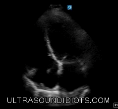
PARASTERNAL LONG AXIS - STANDARD VIEW
-
Start with cardiac/phased array probe
Set machine to cardiac preset
Hold probe like a pencil
Place probe on patient’s left chest, just lateral to the sternum within the 3rd - 5th intercostal spaces.
Orient probe maker to patient’s right shoulder
While watching the screen, start moving the probe from this point slowly spreading out in all directions, making larger movements as you go. Watch the screen the entire time. Watch the heart move in and out of view as you pass over ribs and lung.
Identify the best cardiac window and anchor the probe. Add firm pressure and feel the probe shift into the intercostal space. Stabilize your position by placing your wrist on the patient’s body.
To fully optimize the view, make slow deliberate movements, tilting and rocking the probe in all directions (Mortar and pestle movements)
Focus the mitral value leaflets at the center of the screen and rotate clockwise or counter-clockwise to open up the LVOT.
Max out depth to look for pleural effusion then reduce depth to place descending aorta at the bottom of the screen.
Tips
To help with difficult views, roll patient to left side
If possible, work with patient on breath holding at various points of respiratory cycle
COPD will shift heart inferiorly
PARASTERNAL LONG AXIS - RV INFLOW VIEW
-
Starting from an optimized standard view, tilt probe inferiorly to bring the RV and RA into view.
Rotate clockwise and counterclockwise to optimize view
Tips
If possible, work with patient on breath holding at various points of respiratory cycle.
with small movements, slide the probe in all directions to optimize image.
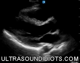
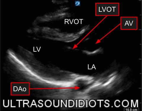
PARASTERNAL LONG AXIS - RV OUTFLOW VIEW
-
Starting from an optimized standard view, tilt probe superiorly to bring the RV and RA into view.
Tips
If possible, work with patient on breath holding at various points of respirator cycle.
with small movements, slide the probe in all directions to optimize image.